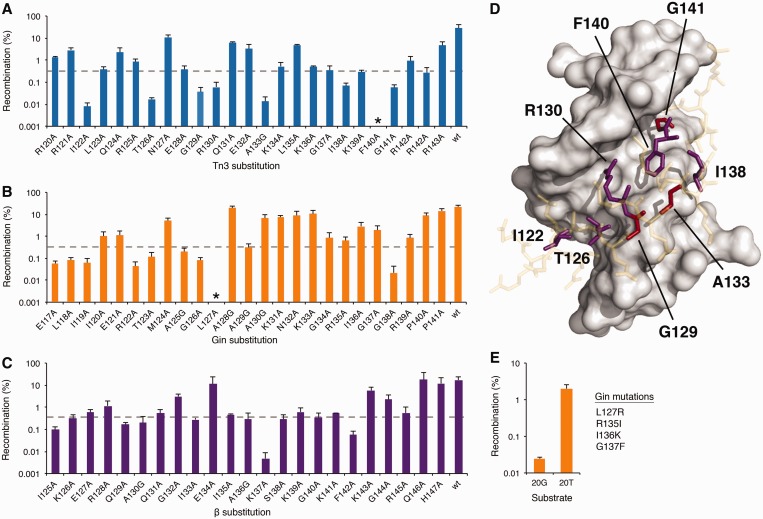
Figure 4.
Alanine-scanning mutagenesis of the serine recombinase arm region. (A–C) Recombination activity of mutant (A) Tn3, (B) Gin and (C) β catalytic domains on their native and minimal DNA targets. Asterisk indicates <0.0001% recombination. Dotted lines indicate threshold below which mutants were considered non-functional. (D) Crystal structure of the γδ resolvase arm region (sticks) in contact with substrate DNA (gray surface). Conserved and variable residues important for recombination are shown in red and purple, respectively. Inert residues are shown in yellow (PDB ID: 1GDT) (20). (E) Recombination by a Gin chimera substituted with residues predicted to impart specificity onto the 20T core site. Recombination was determined by split gene reassembly. Error bars indicate standard deviation (n = 3).

