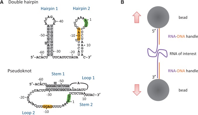
Figure 1.
Schematic drawings of RNA structures and the experimental setup. (A) Two proposed RPSOutr structures, double hairpin (top) and pseudoknot (bottom). The common helices appearing on both conformations are highlighted in grey. The SD sequence (GGAG) and start codon (AUG) are also highlighted. (B) Design of optical tweezers for RNA pulling. RNA of interest was flanked by two handles of RNA–DNA duplexes, which were immobilized on the surface of two polystyrene beads through digoxigenin/anti-digoxigenin antibody (top) and biotin/streptavidin (bottom) interactions. The top bead was trapped by laser beams and the bottom bead was fixed on a micropipette.

