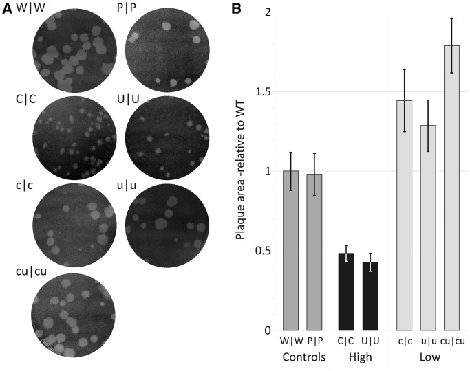
Figure 4.
Plaque morphology of E7 WT and double region mutant viruses. RD cell monolayers in 10-cm plates were infected with a similar infectious titre of virus and incubated for 96 h at 37°C. (A) Plaque appearance. (B) Plaques sizes of WT and mutant E7 viruses calculated from 25 plaques for each virus using ImageJ software (mean values and SEMs relative to WT control shown as bar heights and error bars).

