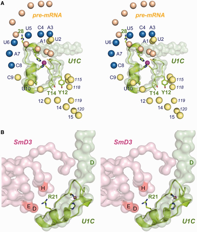
Figure 8.
Disposition of U1C in the structure of the U1 snRNP. (A) Stereo view of the U1C N-terminal domain (light green surface model) and a nearby segment of the U1 snRNA from the 5.5 Å crystal structure of the human U1 snRNP (pdb 3CW1). The NMR structure of U1C-(1-31) (from pdb 2VRD) is superimposed on the crystal structure, with selected side chain depicted as stick models and the Zn2+ ion as a magenta sphere. The phosphate atoms of the U1 snRNA are shown as spheres with the nucleotide numbers specified nearby; the 3ACUUAC8 motif complementary to the 5′SS is in blue. The U1 5′ end base-pairs with its counterpart from an adjacent U1 snRNP complex in the crystal, mimicking the pairing of U1 with nucleotides at the 5′SS of the pre-mRNA intron (colored beige). (B) Stereo view of the U1C (green surface model) and SmD3 (pink surface model) subunits of human U1snRNP, with superimposed NMR structure of U1C, highlighting the proximity of Arg21 (green stick model) to SmD3 residues His11 (H), Glu36 (E) and Asp37 (D). The U1C Asp36 residue is indicated by the green letter D.

