
Figure 2.
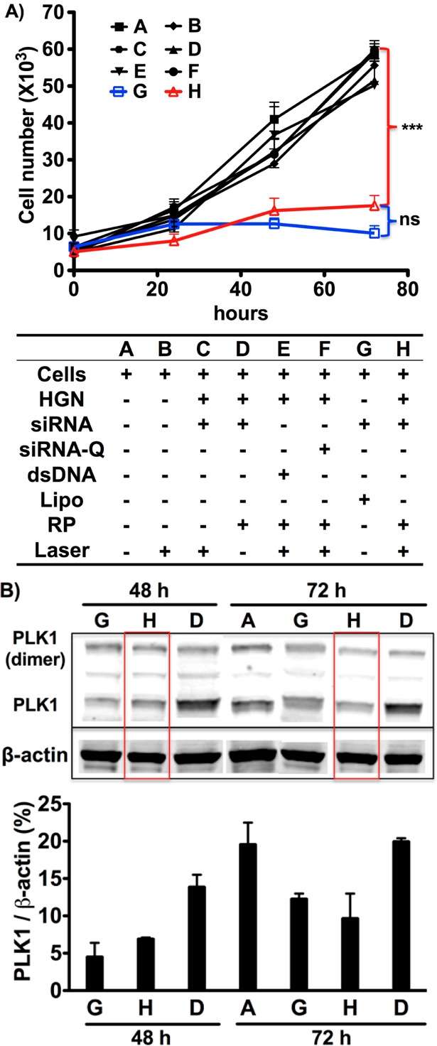
Functional plk1-siRNA released from HGN-SD-RP by NIR-laser (2.4 W/cm2 for 10 s) leads to loss of PPC-1 cell viability and down-regulation of PLK1 protein levels. (A) NIR-laser treatment of PPC-1 cells having internalized HGN-SD-RP (H) causes a significant decrease of cell viability similar to the effect of lipofectamine (G) but at much lower RNAi concentration. A series of controls (defined in the text) are shown in the table underneath the growth curve. ***, p < 0.001; ns, not significant. (B) Western blot analysis showing knockdown of plk1 gene expression in PPC-1 cells. Red boxes highlight the down-regulated expression of plk1 in cells with laser-released siRNA from HGN-SD-RP. The column graph underneath shows the band intensity ratio of PLK1 to β-actin in Western blot image. The HGN-SD-RP provided the similar level of plk1 knockdown as lipofectamine.