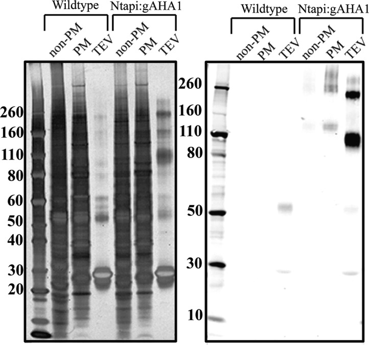
Figure 6.
Purification of Ntapi:AHA1 visulized by western blot. One-hundred micrograms of solubilized plasma membrane protein was incubated with IgG purification resin, washed three times with 0.1% SDS in resuspension buffer followed by three times with AcTEV, and eluted overnight with AcTEV protease. Eluted proteins were acetone-precipitated and resuspended directly in Laemmli sample buffer with reducing agent. One microgram of nonplasma membrane (non-PM) and plasma membrane (PM) wild-type or Ntapi:gAHA1 protein and half of TEV-eluted proteins were loaded on 4–12% NuPAGE gels. The left panel is silver stained for total protein, and the right panel is a western blot probed with rabbit anti-CBP followed by LI-COR IRDye goat anti-rabbit antibody. Size-marker values are in kDa.

