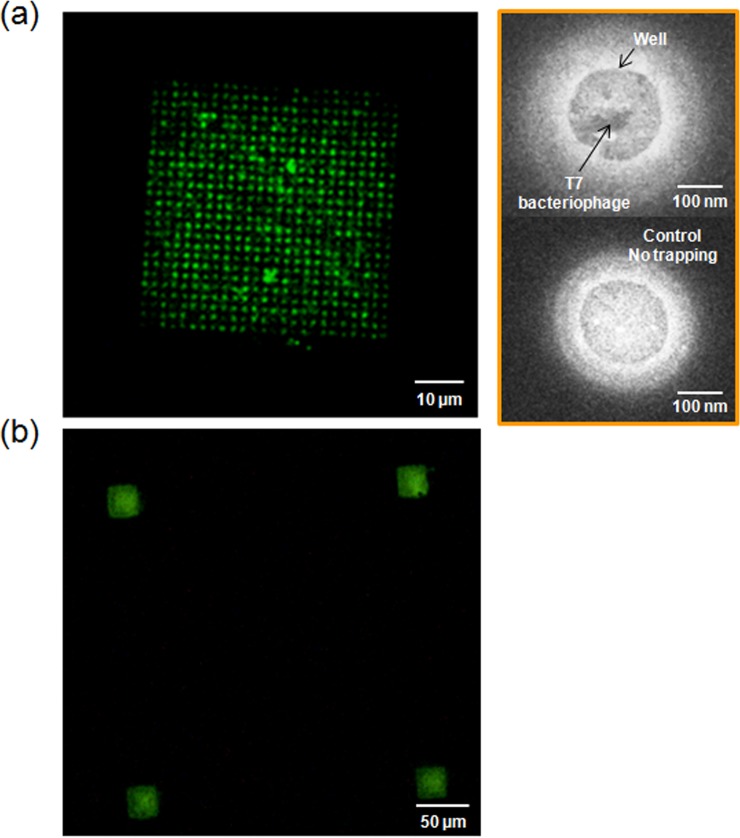
Figure 3.
PC nanostructure arrayed with T7 bacteriophages intercalated with SYBR Green fluorescent dye (excitation: 497 nm, emission: 520 nm). (a) A fluorescent image of 200 nm wells/2 μm periodicity of the array (size of the array: 50 × 50 μm). On the right side, scanning electron microscope images of the T7 bacteriophage trapped into a well (top), In comparison, no bacteriophage trapped well (bottom). (b) A fluorescent image of the arrays with 200 nm wells/350 nm periodicity (size of the array: 25 × 25 μm). The distance between the arrays is 250 μm.

