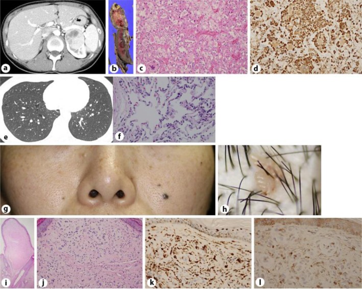
Fig. 1.
The abdominal CT revealed a low-density area on the left kidney of a 37-year-old female with BHD (a), which was histopathologically diagnosed as clear cell carcinoma. On gross examination, the resected kidney contained a partially hemorrhagic, yellowish, solid tumor (b). The tumor was composed of cells with clear to acidophilic cytoplasm (hematoxylin-eosin stain; c), which were positive for vimentin stain (d; 200× magnification). The thoracic CT revealed bilateral, multiple PCs in the basilar and mediastinal regions (e). Microscopic findings for the lung revealed that the septum of the cyst wall contained capillaries and was lined by pneumocytes on both surfaces (f; 200× magnification). Quiet, skin-colored papules were present on the patient's face (g). There were few pinkish-colored, verrucous papules on the scalp (h). Histopathological images (hematoxylin-eosin) of the FF. The stoma was rich in fibroblasts and was oriented in parallel bundles of root sheath-like fibers and neighboring enlarged hair follicles [20× magnification (i) and 200× magnification (j)] positive for factor 13a (k) and c-kit (l; 400× magnification).

