Fig. 3.

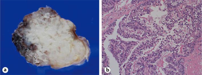
Pathological examination of the intracranial tumor. a Macroscopic findings of the resected tumor. b HE staining of the tumor, ×20.

Pathological examination of the intracranial tumor. a Macroscopic findings of the resected tumor. b HE staining of the tumor, ×20.