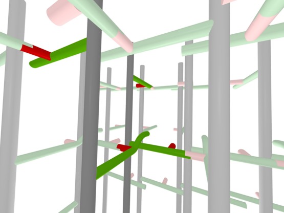
Figure 10.

Three-dimensional representation of the hybrid model for the peptidoglycan lattice of FemA. Two of the glycan chains, peptide stems and glycyl side chains of the central units of the expanded inset (top) of Figure 9 (see red arrows) are highlighted by darker colors. The view is looking from row 3 to row 2 (see Figure 9 for numbering scheme). A cross-linked red glycyl unit is in the foreground (just left of and below the center) as well as an un-cross-linked red glycyl unit (to the right). The steric conflict of the two highlighted green stems in the foreground is represented by an upward curvature of one of the stems.
