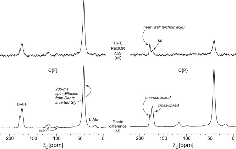
Figure 3.
Dante-selected C{F} (left) and C{P} (right) REDOR of the cell-wall sample of Figure 1. The Dante differencing of Figure 2 preceded REDOR dephasing. Four data blocks were collected resulting in spectra with and without Dante irradiation, each with and without 19F (or 31P) dephasing. The Dante differences (ΔS) are shown at the bottom of the figure and are the reference spectra for REDOR dephasing (ΔΔS) shown above. The terminal carboxyl of the d-alanyl label (178 ppm) has a much larger C{P} REDOR difference than does the peptide d-alanyl label (175 ppm), indicating preferred proximity of un-cross-linked d-alanyl units to wall teichoic acid.

