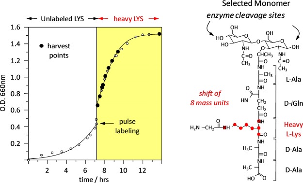
Figure 6.

(Left) Time course of the pulse labeling of FemA whole cells with uniformly 13C,15N-lysine. After the switch to labeled lysine in the media, the cells doubled in about 2 h. (Right) Peptidoglycan digest fragment containing a single labeled lysine.
