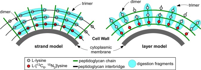
Figure 7.

Muramidase digestion fragments (blue) of pulse labeling of FemA whole cells with l-[13C6,15N2]-lysine (red circles) for the strand model (left), and layer model (right) of peptidoglycan biosynthesis. The fragments with a mixture of heavy and light lysines are only observed in the layer model.
