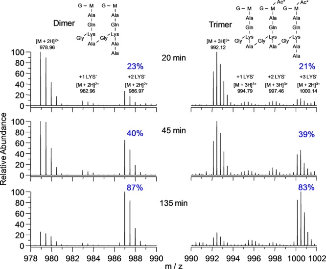
Figure 8.

Accurate-mass spectra of dimers and trimers from digestion of the peptidoglycan of FemA cells grown in the pulse-labeling experiment of Figure 6. The dimers contain 0, 1, or 2 labeled lysines (increasing m/z, left to right), and the trimers, 0, 1, 2, or 3 labeled lysines (left to right), respectively. Each labeled lysine results in a m/z mass shift of 8/2 = 4 units for dimers, and 8/3 = 2.67 units for trimers.
