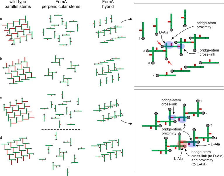
Figure 9.
Lattice models for the peptidoglycan of long-bridged wild-type S. aureus (left) and its short-bridged FemA mutant (middle and right). A 4 × 4 array of glycan chains perpendicular to the plane of the paper is in gray, with the peptide stems in green and the pentaglycyl bridges in red. Panels a through d for each model are slices transverse to the glycan chain direction and separated from one another by a single glycan repeat unit. The model on the left has all nearest-neighbor stems parallel; that in the middle, perpendicular; and that on the right, a mixed geometry. The expanded insets on the far right identify structural components (arrows and color highlights) for the mixed geometry of the FemA hybrid model. The two red arrows (inset, upper right) identify strands that are color highlighted in Figure 10. Alternate rows of the mixed-geometry model have stems that are perpendicular to one another. Thus, the stems of rows 1 and 3 are parallel, and those of rows 2 and 4 are parallel. Counts for the central four strands of the unit cells indicate that ideal cross-linking is 100% for the wild-type lattice (each stem is a cross-link donor and acceptor) and 50% for each of the two FemA models.

