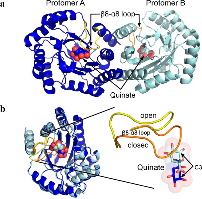
Figure 2.

Distinct conformational behavior within the physiological DHQD homodimer present in the crystallographic asymmetric unit. (a) The DHQD homodimer present in the quinate asymmetric unit is depicted in cartoon representation. Quinate molecules are shown as spheres, and the β8−α8 loops are colored yellow and orange. (b) Superposition of the two protomers highlights the high degree of overall structural similarity but pronounced differences in the β8−α8 loop and quinate conformations.
