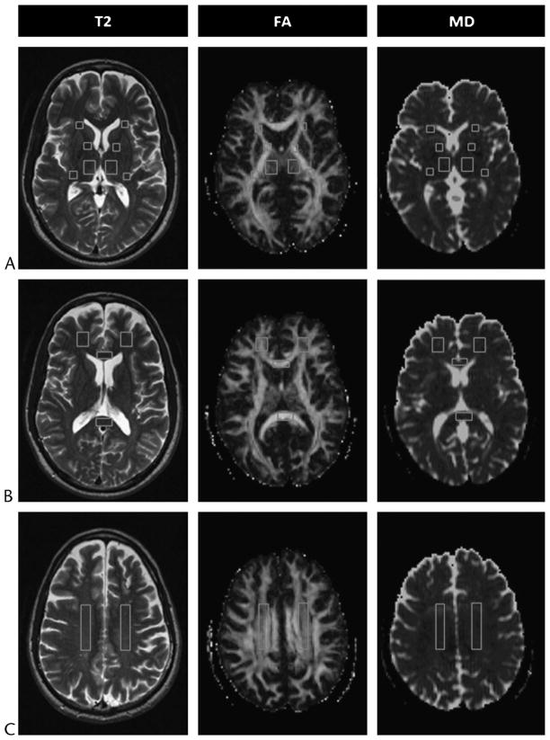
FIGURE 1.
Regions of interest shown on selected axial T2-weighted (T2) images and corresponding FA and MD maps (A, B, and C) from a 49-year-old man with MTBI who showed no visible evidence of brain damage on conventional MRI and was scanned 54 days after injury. Locations of ROIs indicated are as follows: the thalamus and the anterior limb, genu, and posterior limb of the internal capsule (A); the genu and splenium of the corpus callosum and frontal white matter (B); and the centrum semiovale (C). Figure 1 can be viewed online in color at www.topicsinmri.com.

