
FIGURE 4.
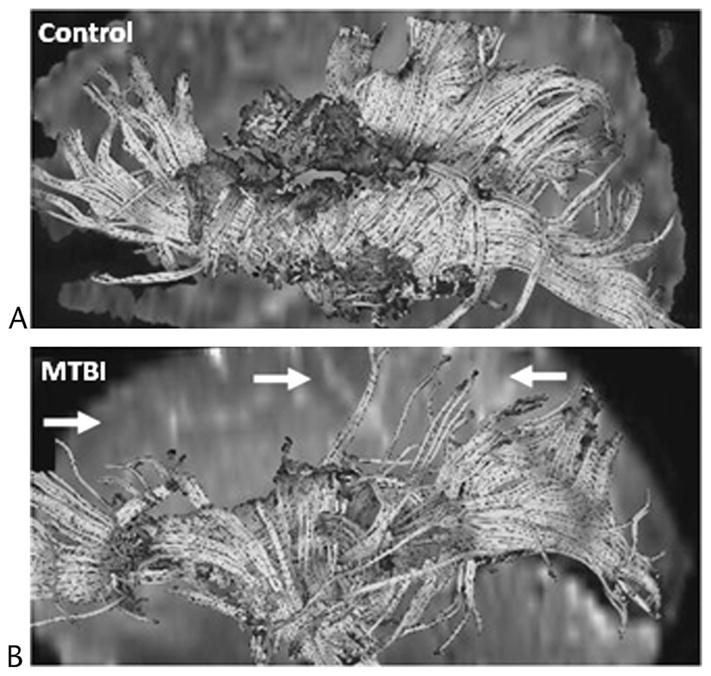
Diffusion tensor imaging tractography results generated from seed voxels placed around the corpus callosum and viewed from the midsagittal region of the left hemisphere for a 56-year-old healthy male control (A) and a 52-year-old male patient with MTBI (B) who showed no visible evidence of brain damage on conventional MRI and was scanned 22 days after injury. In comparison to the control, the patient exhibited fewer frontal, parietal, and occipital white matter fibers tracts (arrows). Figure courtesy of Kelly A. Mcgorty, RT, New York University School of Medicine, New York, NY. Figure 4 can be viewed online in color at www.topicsinmri.com.