Figure 9.
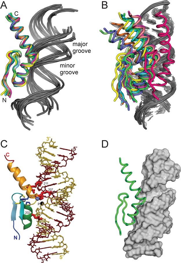
(A, B) Models of the complex between JAZ ZF3 and the minimal dsRNA VAIminMut shown (A) superimposed on the structured portions of the backbone of ZF3 and (B) superimposed on the backbone of the dsRNA duplex. (C) Model of the complex between ZF3 and the minimal dsRNA showing interactions between the side chains of Q202, K207, K208, K211, and Q212 (shown in red) with the phosphate backbone of the RNA. The backbone amide of K207 (blue sphere) and the phosphorus of Cyt13 (black) are shown in close proximity in the complex. (D) Single HADDOCK model with the dsRNA shown in a surface representation, illustrating the docking of the JAZ ZF3 in the major groove. Figure made with (A, B, D) PyMol and (C) Molmol.57

