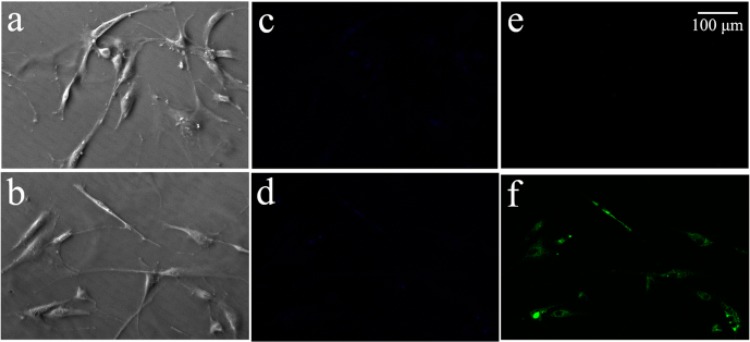
Figure 6.

Fluorescence microscopy images of hMSCs. (a) Bright-field image, and fluorescence images in (c) blue and (e) green channel after hMSCs were pre-treated with 100 μM NEM for 30 min, and then incubated with 20 μM MHF for 1 h. (b) Bright-field image, fluorescence images in (d) blue and (f) green channel after hMSCs being incubated with 20 μM MHF for 1 h at 37°C.
