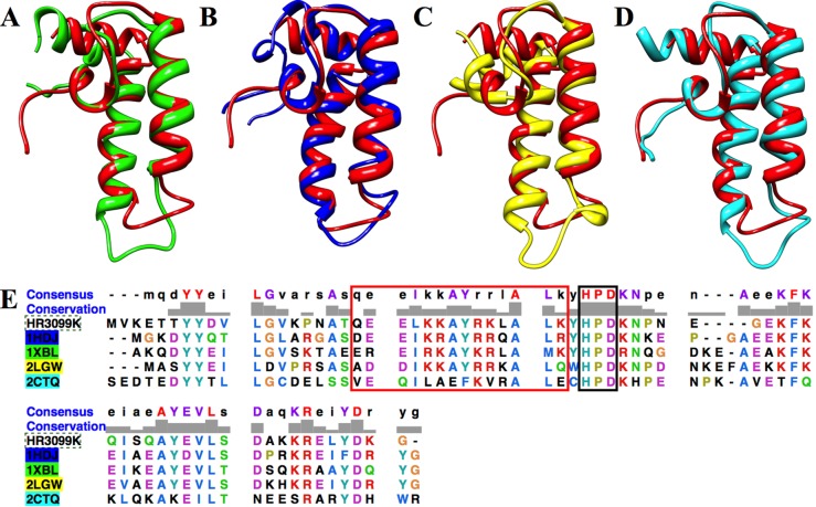
Figure 4.
Overlay of the ribbon structure for DNAJA1-JD (red) with (A) the E. coli DnaJ J-domain (PDB entry 1XBL), (B) the H. sapiens DnaJ homologue subfamily B member 1 J-domain (PDB entry 1HDJ), (C) H. sapiens DnaJ homologue subfamily B member 2 (PDB entry 2LGW), and (D) H. sapiens DnaJ homologue subfamily C member 12 (PDB entry 2CTQ). (E) ClustalW comparison of DNAJA1-JD (HR3099K) with PDB entries 1HDJ (blue), 1XBL (green), 2LGW (yellow), and 2CTQ (cyan). The highly conserved HPD sequence is outlined with a black box. The residues that make up helix α2 are outlined with a red box.

