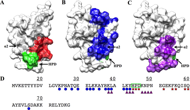
Figure 5.
(A) Transparent surface and ribbon representation of DNAJA1-JD highlighting another proposed DnaK binding site based upon the bovine auxilin–bovine Hsp70 complex (PDB entry 2QWN) (colored red and green; conserved HPD motif). (B) Transparent surface and ribbon representation of DNAJA1-JD (rotated ∼90°) with the proposed DnaK binding site based upon NMR titration data (colored blue). (C) Transparent surface and ribbon of DNAJA1-JD (rotated ∼90°) with the proposed inhibition site based upon the TIM14–TIM16 complex (colored purple). (D) Sequence of DNAJA1-JD with the proposed interaction sites indicated: DnaK binding site from titrations (blue circle), DnaK inhibition site (purple triangles), DnaK binding site from the cross-linked auxilin–Hsp70 complex (red stars), and the highly conserved HPD motif (green box). The conserved HPD motif and helix α2, which is potentially an important component of the DnaJ–DnaK interaction site and the TIM16-like inhibitory binding site, are labeled.

