
Figure 3.
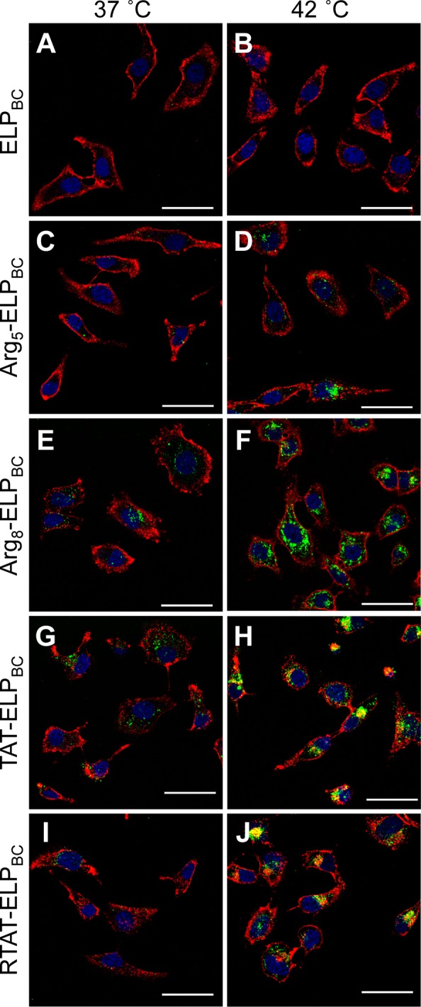
Visualization of controlled cellular uptake of CPP-ELPBCs by confocal fluorescence microscopy. HeLa cells were incubated for 1 h at 37 or 42 °C with 15 μM ELPBC (A,B), Arg5-ELPBC (C,D), Arg8-ELPBC (E,F), TAT-ELPBC (G,H), or RTAT-ELPBC (I,J). Cells were briefly incubated with Alexa 594 wheat agglutinin and Hoechst 33342 to stain the cell membrane and cell nuclei, respectively. At 42 °C, the CPP-ELPBCs self-assembled into micelles displaying enhanced CPP density on the micelle corona, which resulted in amplified cellular uptake, though to differing extents, for the CPP-ELPBCs, as compared to 37 °C, at which all CPP-ELPBCs existed as soluble unimers presenting only a single CPP per polypeptide. In contrast, the nonfunctionalized ELPBC control showed no difference in cellular uptake between 37 and 42 °C. Red, cell membrane; blue, cell nuclei; green, ELPBC; scale bars, 50 μm.