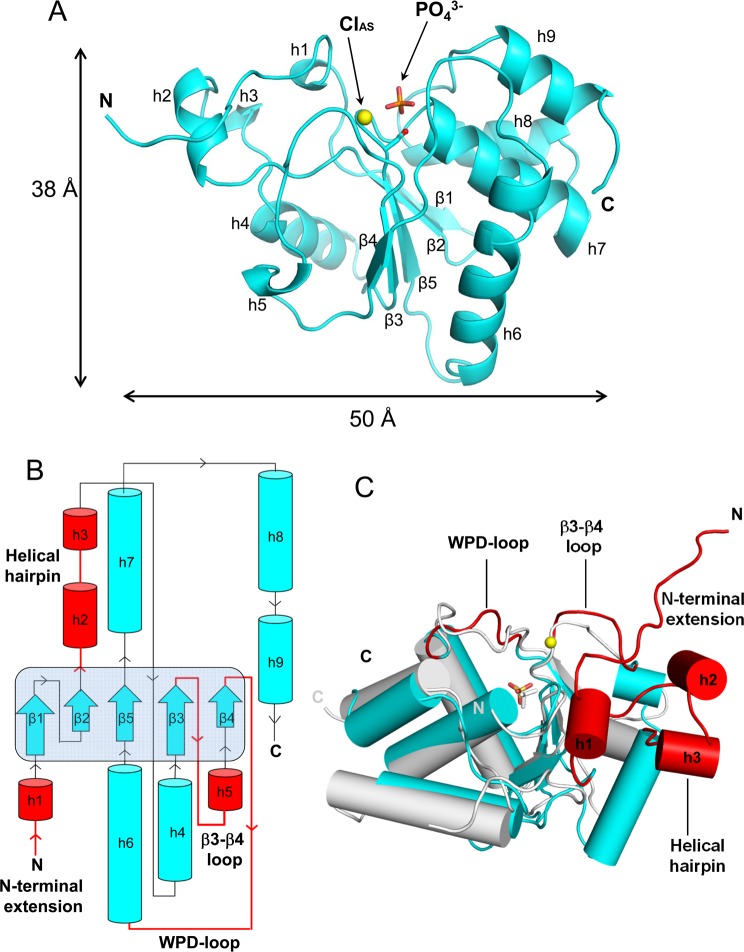
Figure 2.
Atomic structure of PIR1-C152S-core determined at 1.20 Å resolution. (A) Ribbon diagram of PIR1-C152S-core with S152 shown as a red sphere. The active site phosphate and chloride ions are colored orange and yellow, respectively. (B) Topological diagram of PIR1-C152S-core. The central β-sheet formed by strands β1−β5 is highlighted with a light blue background. Insertion elements not found in VH1 are colored red. (C) Superimposition of PIR1-S152C-core (cyan) and VH1 (gray) with helices shown as cylinders (for the sake of clarity, the VH1 N-terminal helix spanning residues 1–20 was omitted). Colored red are the structural elements that render the PIR1 catalytic cleft significantly deeper than in VH1.

