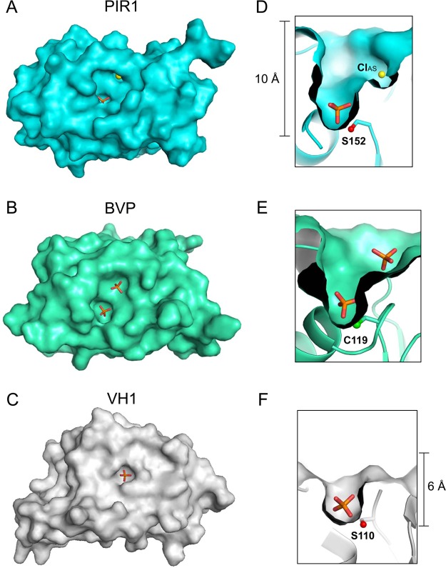
Figure 3.
Depth of the PIR1 catalytic cleft. Surface representations of (A) human PIR1-C152S-core, (B) baculovirus BVP (PDB entry 1YN9), and (C) Vaccinia virus monomeric VH1 (PDB entry 3CM3) reveal striking differences in the width of the active site pocket. Magnified cut-through views of (D) PIR1, (E) BVP, and (F) VH1 catalytic pockets reveal differences in the depth of the active site crevice.

