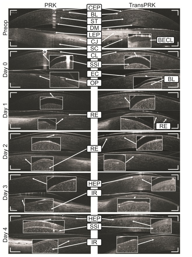
Fig. 3.
SOCT tomograms of the central cornea and peripheral cornea with limbus, acquired before the surgery and in the first postoperative day after PRK and TransPRK. Legend: CEP – corneal epithelium, BECL – basal epithelial cell layer, BL – Bowman’s layer, ST – stroma, DM – Descemet’s membrane, LEP – limbal epithelium, CJ – conjunctiva, SC – sclera, CL – contact lens, OP – opaque epithelium, RE – regrown epithelium, EC – cut of the epithelium, IR – inflammatory response, HEP – hyperreflective epithelium, SSI – stromal surface irregularities, (stars) – artifacts caused by specular reflection. The preoperative refractive error of the eye after PRK was: −2.5Dsph −2.0Dcyl ax 172°, after TransPRK: −4.75Dsph −0.5Dcyl ax 80°. Scale bars in both direction represent 500 µm.

