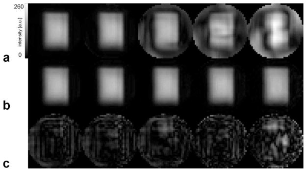
FIG. 1.

a: Spectroscopic water image of spherical phantom filled with a solution of brain metabolites at physiological concentration levels from a dataset acquired without water suppression as reconstructed by gridding reconstruction for acceleration factors 1, 2, 3, 4, and 6 (left to right). The PRESS module selected a 9 × 13 × 2-cm3 box through the center of the sphere. b: Same as (a) but data reconstructed with iterative SENSE. c: Difference between the images in (b) and the water image reconstructed with the reference standard (scaled up by factor of 10).
