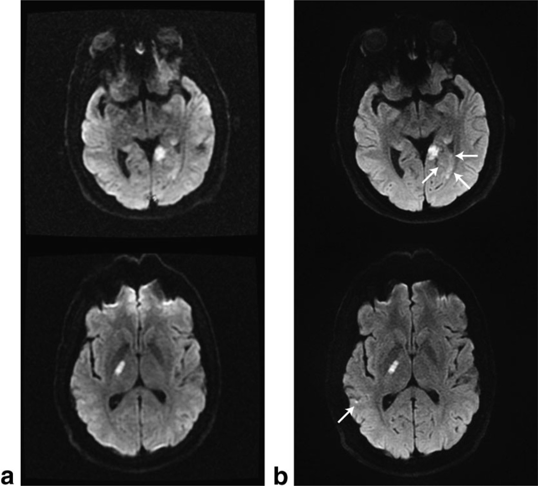
FIG. 6.
Clinical examples of the standard diffusion sequence using (a) 128 × 128 ss-EPI data acquired in less than 1 min, and (b) our three-shot 192 × 192 DW-EPI data reconstructed with GRAPPA using a tetrahedral diffusion scheme repeated three times. The top and bottom rows were acquired with the eight-channel neurovascular array coil and the eight-channel brain coil, respectively.

