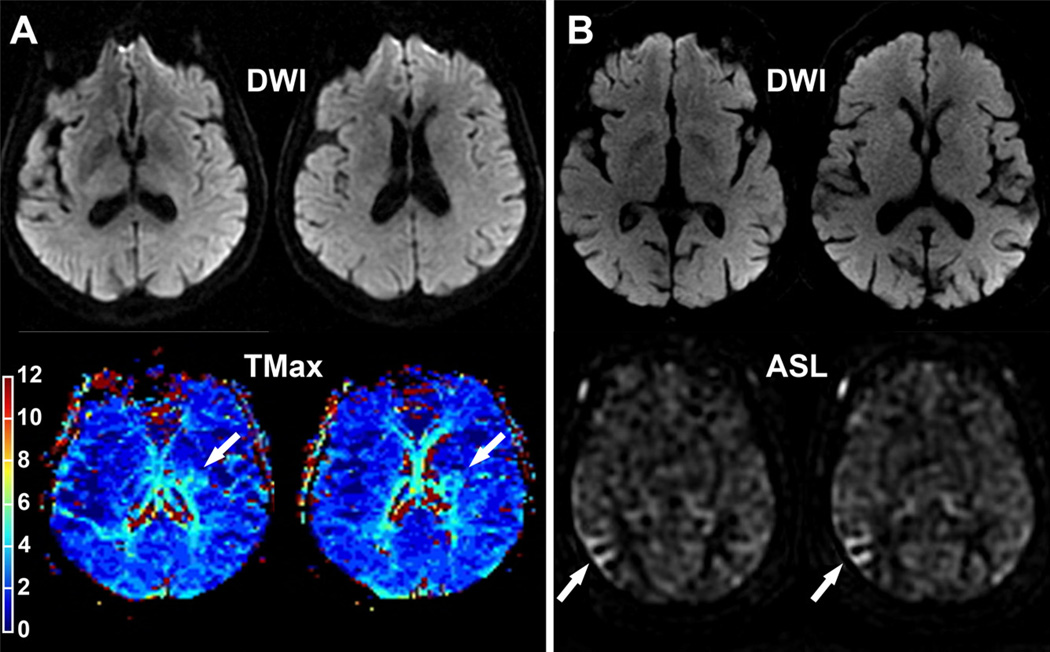
Figure 2.
A, A 75-year-old patient referred for a transient episode of right-sided weakness (ABCD2=4). MRI was performed 5 hours after symptom onset. Diffusion-weighted sequence was negative. TMax revealed a perfusion lesion in the deep left MCA territory (arrow). This lesion progressed into infarction 4 days later when the patient developed a persistent right-sided weakness. B, An 82-year-old right-handed patient with atrial fibrillation referred for a transient episode of left-sided weakness. ABCD2 score=6. MRI was performed 22 hours after symptom onset. Acute DWI was negative. ASL identified hypoperfusion in the right MCA territory (arrow). TMax the time when the residue function reaches its maximum; MCA, middle cerebral artery; DWI, diffusion-weighted imaging; ASL, arterial spin labeling.

