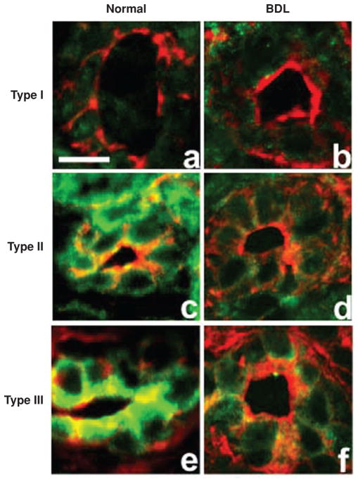
Figure 4.
Inositol 1,4,5-trisphosphate receptors (InsP3R) expression is lost in bile duct epithelia after bile duct ligation. Confocal immunofluorescence of liver sections from normal rats and rats subjected to bile duct ligation (BDL) labeled with isoform-specific InsP3R antibodies (green) and rhodamine phalloidin (red). Type 1 InsP3R labeling in normal bile duct cells (A) is found throughout each cell although is expressed at low levels, similar to that observed 2 weeks after BDL (B). Type 2 InsP3R labeling is seen throughout each cell in normal liver sections (C) and it is nearly absent 2 weeks after BDL (D). Type 3 InsP3R labeling is found predominantly in the apical region of bile duct cells in normal liver sections (E) and is also markedly reduced 2 weeks after BDL (F) [reprinted from reference (354), with permission].

