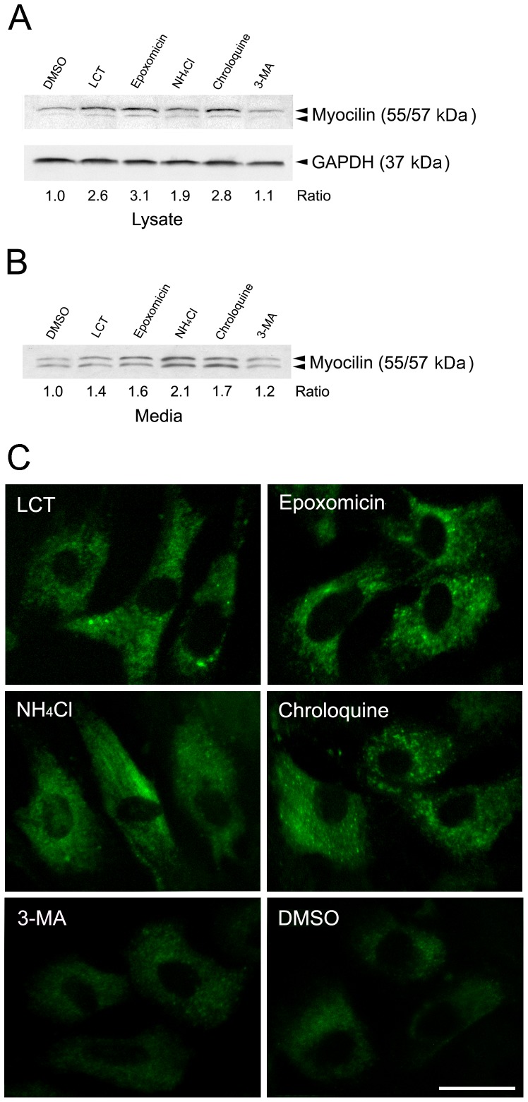
Figure 2. Effects of inhibitors on levels of endogenous myocilin.
A. Western blotting. Human TM cells were treated for 16 h with vehicle DMSO or H2O (not shown), or proteasomal (LCT and epoxomicin), lysosomal (NH4Cl and chloroquin) or autophagic (3-MA) inhibitors. Proteins (25 µg) in cell lysates were immunoblotted with anti-myocilin or anti-GAPDH. Densitometry was performed. The myocilin/GAPDH relative to the DMSO control ratios are presented. B. Equal aliquots of media were immunoblotted with anti-myocilin. The level of myocilin in the media was normalized to that of the DMSO control. C. Immunofluorescence for myocilin. TM cells treated as above with the various inhibitors were fixed and immunostained for myocilin. All experiments were repeated at least 3 times, yielding similar results. Scale bar, 20 µm.

