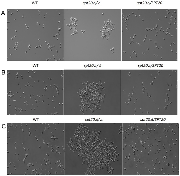
Figure 5. Micrographs of C. albicans morphology.
Cell morphology was examined. Micrographs after 2(A), 8 h (B), and 24 h (C) of growth at 30°C are presented for the wild-type, spt20Δ/Δ and spt20Δ/SPT20 reintegrated strain. The wild-type and spt20Δ/SPT20 re-integrated cells separate and appear to have a round morphology at all 3 time points while spt20Δ/Δ cells cluster together and demonstrate a “snow-flake” shape.

