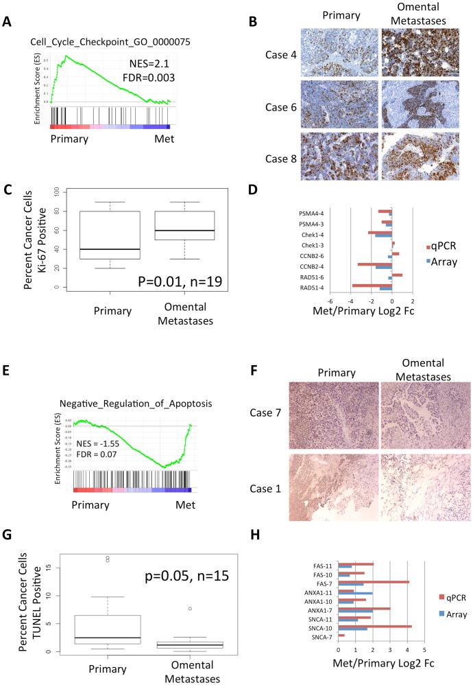
Figure 1. Metastases are more proliferative and include less apoptotic cells than matched primary tumors.
A. GSEA enrichment plot suggests Reactome G2/M cell cycle checkpoints including Chek1, CCNB2, and BUB1 are expressed higher in primary tumors. B. Representative immunohistochemical staining of Ki-67 from three cases suggest higher Ki-67 staining in omental metastases. C. Box plot of the percent cells with positive Ki-67 staining. Paired t-test suggests significant differences between omental metastases and primary tumors from 19 cases. D. qPCR validation of cell cycle checkpoints. Note that genes with significant >1.8 fold changes in the array validate in those same tumors by qPCR. Genes with small expression changes measured by either the array or qPCR may be noisy in those tumors. E. GSEA enrichment plot suggests that negative regulators of apoptosis are up-regulated in metastases. F. TUNEL staining of two representative cases shows increased TUNEL signal in metastases. G. Box plot of the percent of cancer cells as determined by H&E staining with positive TUNEL staining. Paired t-test suggests significance.

