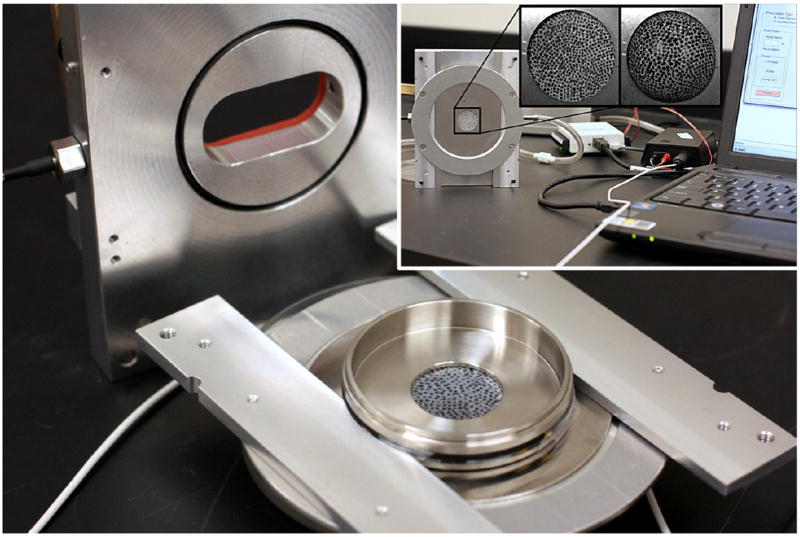
Figure 1.

Main components of the cell stretching device: 1) a top plate with glass window, pressure probe and gas inlet: 2) a bottom plate and 3) a round stainless steel cell cultured plate with a circular opening (diameter: 20 mm). Inset: compressed air is introduced into an airtight chamber, which results in deformation of the PDMS membrane, and thus stretching the adherent cells. The PDMS with stochastic pattern was used only in strain calibration experiments (details are described elsewhere (Skotak, Wang, 2012)).
