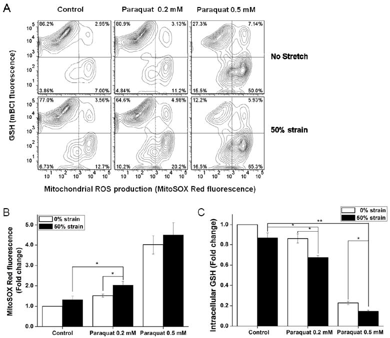
Figure 6. Effect of stretch on mitochondrial ROS and intracellular GSH levels in SH-SY5Y cells treated with paraquat.

Cells were stretched at 50% strain 2 h before exposure to paraquat. MitoSOX and mBCl fluorescence was evaluated 48 h after paraquat treatment by FACS. A. Representative histograms depict the changes in MitoSOX and mBCl fluorescence in response to the indicated treatment. In B and C, data are expressed as mitochondrial ROS production (MitoSOX Red fluorescence) normalized against control values. Data in graphs represent mean ± SEM of n = 5 experiments. *P< 0.05 and **P< 0.01.
