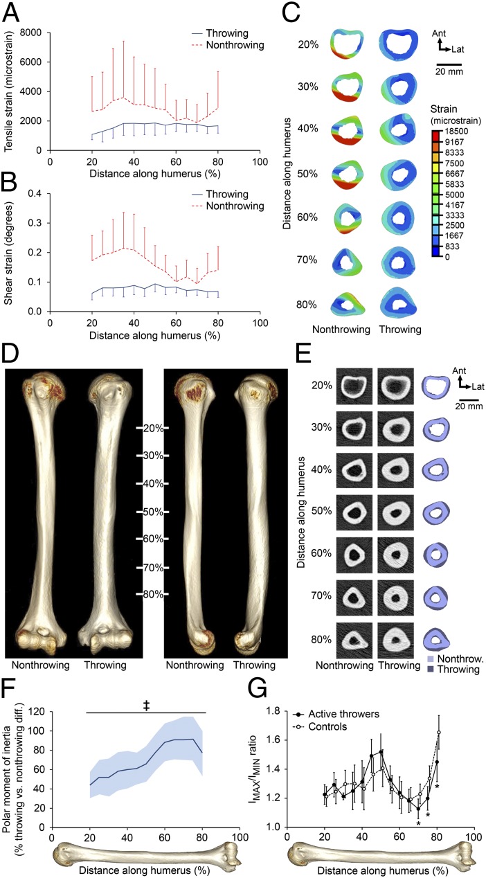
Fig. 1.
Overhand throwing loads the humeral diaphysis, inducing skeletal adaptation. (A and B) Median (and median absolute deviation) cross-sectional tensile (A) and shear (B) strains in the humerus of the throwing arm of an MLB player toward the end of the cocking stage of a fastball pitch showed strains throughout the diaphysis. Both tensile and shear strains were increased when the same forces were applied to the collateral, nonthrowing humerus. (C) Cross-sectional distribution of peak tensile strain in the bilateral humerus demonstrated reduced strains in the throwing arm. (D) Reconstructed CT images of the bilateral humerii in a representative MLB/MiLB player demonstrated a more robust diaphysis with visibly broader diameter on the throwing side. (E) Cross-sectional images of the humerii in D revealed substantially greater total and cortical bone areas and cortical thickness and smaller medullary area in the throwing arm. (F) Throwing substantially increased torsional bone strength (indicated by density-weighted polar moment of inertia) along the entire diaphysis, with strength nearly doubled toward the distal diaphysis. Data show the mean percent difference and 95% CI (shaded area) between the throwing arm and the nonthrowing arm in throwers normalized to the differences between the dominant arm and the nondominant arm in controls (‡P < 0.001, unpaired t test). (G) The distal diaphysis in throwers had a more circular cross-section than seen in controls, as indicated by a maximum:minimum (IMAX:IMIN) second moment of area ratio closer to 1 (*P < 0.05, ANCOVA with the contralateral arm as the covariate).

