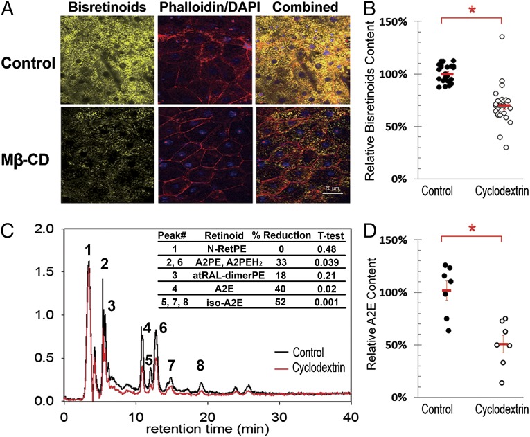
Fig. 5.
Mβ-CD removes LBs from Abca4-Rdh8 DKO eyes ex vivo. (A) Flat mounted eyecups from Abca4-Rdh8 mice after 72 h incubation in culture media alone (control) or supplemented with 500 μM Mβ-CD. RPE borders and nuclei were labeled with phalloidin (red) and DAPI (blue), respectively. Auto fluorescent LB granules are in yellow. (B) Quantification of lipofuscin’s mean fluorescence intensity per randomly chosen field in controls (n = 3) and Mβ-CD treated (n = 3) eyecups. Means ± SEM of the three experiments are in red. The attained reduction in bisretinoid fluorescence was 31% (P < 0.0007). (C) Representative HPLC chromatogram showing the bisretinoids profile of control (black trace) and Mβ-CD-treated (red trace) eyecup extracts. The inserted table depicts the average reduction of the areas under the peaks for all identified retinoids (n = 7 control eyes and 7 treated eyes). All identified peaks show reduction. In addition, there are some minor unidentified peaks that also show reduction; they likely correspond to small amounts of oxidized bisretinoids. (D) Quantitation by HPLC of total A2E. Individual dots represent the total content of A2E (A2E plus iso-A2E) per eyecup. Seven age-matched eyecup extracts from Abca4-Rdh8 DKO mice for each group, control (black) and Mβ-CD-treated (open) are presented in the graph. The average content and SE for controls and Mβ-CD treated eyecups are indicated in red. The difference corresponds to a reduction of 48% (P < 0.005).

