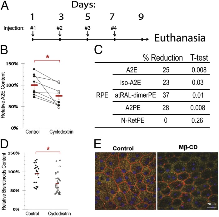
Fig. 6.
Intravitreal Mβ-CD reduces LB in the RPE of Abca4-Rdh8 DKO mice. (A) Schematic diagram of intravitreal injections and euthanasia. Right (treated) and left (controls) eyes received 1.5 µL of 100 mM Mβ-CD in PBS and PBS, respectively, in each administration. Eye specimens were analyzed by HPLC and Microscopy. (B) Difference in the content of total A2E (A2E and iso-A2E) between left (control) and right (cyclodextrin) eyes determined by HPLC. Each dot represents one eye (n = 8). Red symbols are average content and SEs. The difference between eyes corresponds to a reduction of 24% (P < 0.013). (C) Differential content of bisretinoids and bisretinoid precursors in the RPE as determined by HPLC. (D) Quantification of the content of bisretinoids in the left and right eyes by quantitative fluorescence microscopy. The difference between eyes corresponds to 27% (P < 0.01). (E) Representative inmmunofluorescence of flat mounted eyecups depicting the integrity of the RPE layer. CD treatment reduced the number and fluorescent intensity of lipofuscin granules. Actin filaments at cell borders (red) and nuclei (blue) were decorated with phalloidin and DAPI, respectively. All panels are at the same magnification (×400).

