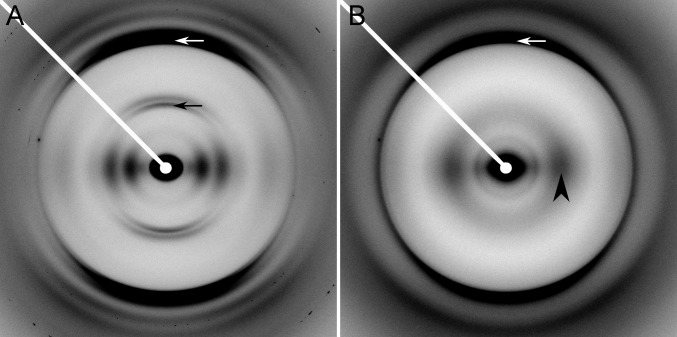
Fig. 2.
Representative X-ray fiber diffraction patterns. (A) HET-s(218–289) WT showing two-rung β-solenoid diffraction. (B) HET-s(218–289) mutant RL showing characteristic stacked β-sheet diffraction. White arrows, 4.7-Å meridional reflection; black arrow, 9.4-Å meridional reflection; and black arrowhead, equatorial ∼10-Å intensity maximum corresponding to the intersheet spacing.

