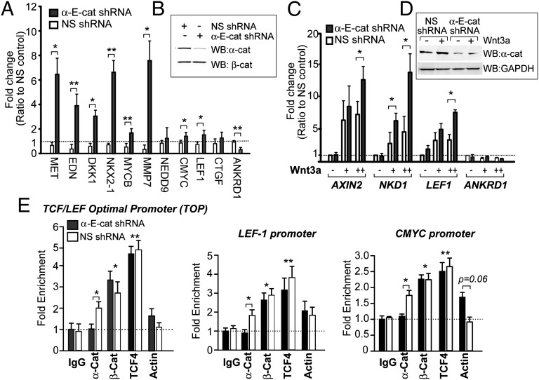
Fig. 2.
α-Cat inhibits β-cat signaling. (A) qPCR of β-cat target genes from SW480 cells stably transfected with nonspecific (NS) and α-E-cat shRNAs and (B) the corresponding immunoblot. (C) qPCR of β-cat target genes from human dermal fibroblasts stably transfected with NS and α-E-cat shRNAs and incubated ±Wnt3a. Genes in A and C are normalized to 18S and fold induction was calculated with the ΔΔCt method from three independent experiments. (D) Corresponding immunoblot. (E) ChIP analysis of α-cat, β-cat, TCF4, and actin enrichment at three established β-cat/TCF–responsive promoters in nonsilenced and α-cat–silenced SW480 cells: integrated 4xTOP promoter, LEF1, and C-MYC. Asterisks denote significance by standard t test (*P < 0.05 and **P < 0.01). Those without brackets correspond to both silenced and nonsilenced cells compared with the IgG control. Error bars represent SEM for 2–3 independent experiments.

