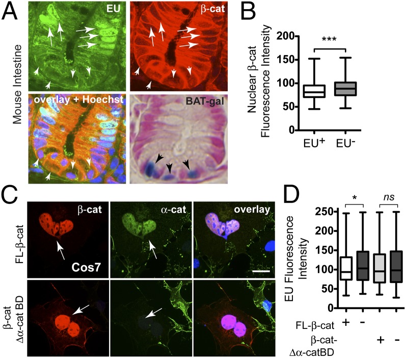
Fig. 6.
β-Cat attenuates transcription by its α-cat–binding domain. (A) Intestinal crypt image from a EU-labeled mouse (2 mg; 4 h) and RNA detection (green) by alkyne–azide click chemistry (SI Appendix, Materials and Methods) and double labeled with an antibody to β-cat (red). Nuclear β-cat+ stem cells at crypt base (arrowheads) incorporate less EU than nearby transit amplifying cells (arrows). X-gal staining (blue) of crypts from β-cat/TCF reporter mouse (BAT-gal; Jackson Labs) shows peak reporter activity in basal (stem) cells. Images taken with 63× objective. (B) Quantification of nuclear β-cat mean fluorescence intensity in EU+ and EU− cells (n = 276; ***P < 0.002). (C) Image of COS7 cells transfected with full-length β-cat– and β-cat-Δα-cat–binding domain and labeled with antibodies to β-cat (red), endogenous α-cat (green), and DNA (blue). (D) EU labeling of cells transfected with β-cat or β-cat lacking the α-cat–binding domain (β-cat-Δα-cat BD). Mean fluorescence intensities from 200 transfected (+) and adjacent untransfected (−) cells were quantified in MetaMorph (*P < 0.05 by t test; see also SI Appendix, Fig. S15). ns, not significant. Arrows point to transfected cells; error bars represent SEM. (Scale bar: 10 μm.)

