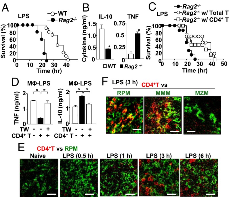
Fig. 1.
CD4+ T cells protect mice from LPS endotoxemia and suppress macrophage TNF production via interaction with macrophages. (A) Endotoxemia induced by i.p. LPS (40 mg/kg mouse weight) injection in WT mice (n = 10), Rag2−/− (n = 5). (B) Serum levels of IL-10 and TNF in mice at 17 h after LPS injection. (C) LPS endotoxemia in T-cell–reconstituted Rag2−/− mice. Data were obtained from Rag2−/− (n = 8), T-cell–reconstituted Rag2−/− (n = 6), and CD4+ T-cell–reconstituted Rag2−/− mice (n = 7). (D) Macrophages were cultured in the medium containing LPS (100 ng/mL) for 24 h. CD4+ T cells were added either directly or separately with a transwell (TW) insert as indicated. (E) Localization of CD4+ T cells (red) and red-pulp macrophages (RPM) (F4/80) (green) in the spleen isolated from naive mice and LPS-treated mice at indicated time points. Staining to localize CD4+ T cells and RPM is shown with red and green, respectively. (Scale bar, 25 μm.) (F) Localization of CD4+ T cells (red) and subsets of macrophages (green); RPM (F4/80), marginal metallophilic macrophages (MMM) (MOMA-1), or marginal zone macrophages (MZM) (SIGN-R1) in the spleen from mice 3 h after LPS treatment. (Scale bar, 15 μm.) All of the experiments are representatives from at least two similar experiments for each. *P < 0.05.

