
Fig. 2.
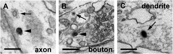
Examples of unlabeled and labeled coated vesicles. (A) An unlabeled coated vesicle (arrow) inside an axon of a specimen that was fixed 10 s after stimulation. The arrowhead points at a noncoated labeled vesicle. (B) A labeled coated vesicle (arrow) and a labeled large vesicle (arrowhead) inside a presynaptic bouton fixed 20 s after stimulation. Note that both labeled vesicles are outside the main SV cluster to the left. (C) A labeled coated vesicle (arrow) inside a dendrite. (Scale bars: 100 nm in A, 300 nm in B, and 200 nm in C.)