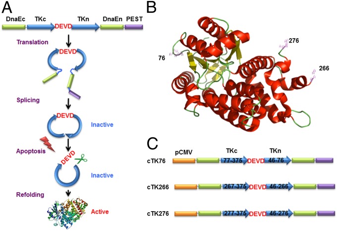
Fig. 1.
The cTK reporter probe for apoptosis imaging. (A) Schematic overview of the principle for monitoring apoptosis. The N and C termini of HSV1-TK are linked with DEVD, a substrate peptide of caspase-3. Upon caspase-3 activation during apoptosis, the DEVD sequence is cleaved and the cyclized TK restores its activity. (B) The crystal structure of truncated HSV1-TK with 1–45 aa deletion. The subunits of TK display a general αβ folding pattern, each of which consists of 13 α-helices (in red) and seven β-sheets (in yellow). The numbers represent the split sites. (C) Schematic structure of the variants of cTK constructs. Each construct is made by cloning into a pcDNA3.1 (+) plasmid backbone, under the control of a CMV promoter.

