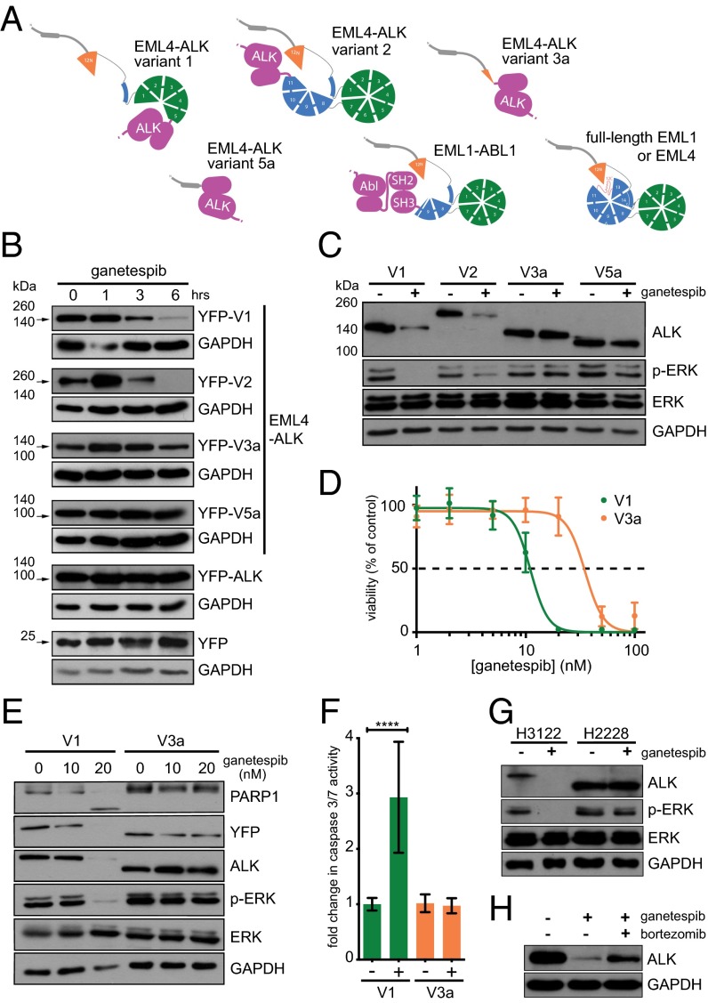
Fig. 3.
The TAPE domain structure explains differential stability of EML4-ALK variants in response to Hsp90 inhibition. (A) Models of full-length EML1 and EML4 and EML1-ABL1 and EML4-ALK fusion variants based upon the EML1 TAPE domain structure are illustrated schematically. (B) MEFs were transfected with YFP, YFP-cytoplasmic ALK (amino acids 1,058–1,620: the region of ALK present in the fusion proteins), or YFP-EML4-ALK variants 1, 2, 3a, and 5a. Forty-eight hours post transfection, cells were treated with 100 nM ganetespib for a further 1, 3, and 6 h. Transient overexpression of EML4-ALK was assessed by Western blot with anti-GFP 4B10 antibody as indicated by arrows. (C) Ba/F3 clones stably expressing YFP-EML4-ALK variants 1, 2, 3a, or 5a were left untreated or treated with 20 nM ganetespib for 6 h. Expression of ALK, ERK phosphorylated at T202 and Y204 (p-ERK), and total ERK was determined by Western blot. GAPDH was used as a loading control. (D) Ba/F3 clones expressing V1 or V3a were left untreated or treated with ganetespib doses ranging from 1 to 100 nM. Cell viability was assayed after 24 h of treatment and normalized as the percentage of the untreated control for each cell line. (E) Ba/F3 clones stably expressing variants 1 or 3a were left untreated or treated with 10 or 20 nM ganetespib for 12 h. Expression of ALK, ERK phosphorylated at T202 and Y204 (p-ERK), and total ERK were determined by Western blot. Induction of apoptosis was analyzed with anti-PARP1 antibody. GAPDH was used as a loading control. (F) V1 and V3a clones were left untreated or treated with 20 nM ganetespib for 12 h and assayed for caspase 3/7 activity. (P values: V1 < 0.0001; V3a = 0.4086). (G) NCI-H3122 and NCI-H2228 (NSCLC cells endogenously expressing EML4-ALK variant 1 and 3b, respectively) were untreated or treated with 100 nM ganetespib for 6 h and analyzed by Western blot as in C. (H) NCI-H3122 cells were left untreated or treated with 100 nM ganetespib alone or in combination with 100 nM bortezomib for 6 h. ALK expression was determined by Western blot with anti-ALK.

