
Fig. 2.
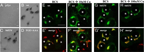
wtATP7B and ATP7BS653Y have similar Cu(I) transport activity yet show different trafficking behaviors in polarized hepatic cells. (C) ATP7BS653Y exhibits Cu(I) transport activity. YST cells were cotransfected with pTyrosinase (pTyr) and the indicated plasmid as described in Methods. The black reaction product reflects copper-dependent tyrosinase activity. Negative controls (A and D) and wtATP7B (B) were always included. (Magnification: A–D, 40×.) WIF-B cells were infected with wtATP7B and ATP7BS653Y adenovirus, cultured overnight in 10 μM BCS, then kept in the chelator (E, E′, G, and G′), or incubated in 10 μM CuCl2 (F and F′) or 100 μM CuCl2 (H and H′) for 1 h, fixed, stained with antibodies to GFP (green in E, F, G, and H) and TGN38 (red in E′, F′, G′, and H′), and imaged by confocal microscopy. Single optical sections are shown. Exogenous wtATP7B redistributes from the TGN (E and E′) to vesicles and the apical membrane when copper levels are elevated (F and F′). In contrast, ATP7BS653Y remains in the TGN in the absence (G and G′) or presence of copper (H and H′). n, nucleus; *, apical space. (Scale bars, 10 μm.)