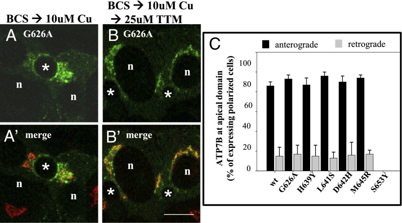
Fig. 3.
Qualitative and quantitative evidence that five WD patient mutations show normal trafficking but ATP7BS653Y does not. (A and B) WIF-B cells infected with adenoviruses encoding wtATP7B and each of the patient mutations were processed for anterograde and retrograde trafficking as described in Methods. (A and A′) In the presence of copper, GFP-ATP7BG626A fluorescence is seen at the apical membrane and in vesicles; it does not overlap with the TGN marker (red). (B and B′) After copper chelation, the protein is no longer seen at the apical membrane and overlaps strongly with the TGN marker. (C) The numbers of polar cells with ATP7B protein fluorescence at the apical surface in the presence and after copper chelation were determined and expressed as a percentage of the total polarized cells expressing the ATP7B protein. Five of the mutant ATP7B showed wild-type trafficking in copper and after copper chelation. However, the ATP7BS653Y mutant was not found at the apical surface under any copper condition; it was not evaluated using the retrograde assay. The numbers of polar cells evaluated for anterograde/retrograde trafficking, respectively, were: G626A: 310/152; H639Y: 193/172; L641: 202/118; D642H: 242/168; M645R: 228/123; and S653Y: 228. (Scale bar, 10 μm.)

