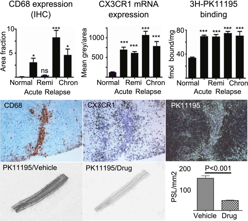
Fig. 3.
AZD8797 treatment in EAE reduces TSPO binding, a clinically relevant marker for microglia activation. (Top) Quantification of CD68 immunoreactivity, CX3CR1 mRNA (in situ hybridization), and TSPO receptor autoradiography ([3H]PK11195) on tissue sections from the lumbar spinal cord of rats during different phases of EAE; n = 33 rats with EAE (10 acute, 8 remi, 10 relapse, 5 chron) and 9 controls. chron, chronic phase; remi, remission. (Middle) Representative 20× photos of adjacent sections of a perivascular EAE lesion for each of the three markers (CD68, bright field; CX3CR1 bright field; PK11195, dark field). In this plaque, the CD68+ (brown label) cells were most significantly located at a perivascular position, whereas the CX3CR1+ (black silver grain) cells were distributed within the surrounding parenchyma. PK11195 autoradiography (bright silver grains) detected both of these population of cells. (Bottom) Bar graphs illustrates significant treatment effect of AZD8797 at 81.8 µmol/kg per day (from day 10–26 post-EAE induction) on TSPO binding, measured with receptor autoradiography of 1 nM [3H]PK11195. Pictures show representative tissue sections from lumbar spinal cord of AZD8797 or vehicle treated EAE rats (n = 20 rats per group).

