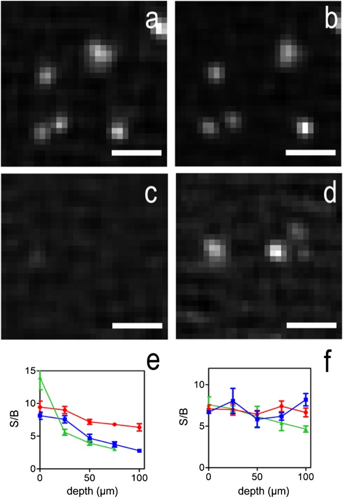
Fig. 2.
One- and two-photon MSIM imaging at different depths in scattering artificial samples. Fluorescent beads (yellow-green 0.1 µm) suspended in 3% agarose gel were imaged as a function of depth and scattering. The imaging laser powers were chosen to give similar fluorescence between the two imaging modes in the absence of scattering beads. Images were collected in 5-µm volumes on the same fields of beads for each imaging mode at depths of 0, 25, 50, 75, and 100 µm with the addition of 0%, 0.13%, or 0.26% polystyrene nonfluorescent scattering beads. In the absence of scattering beads at 50-µm depth, beads are visible in both (A) 1P- (B) and 2P-MSIM modes. In contrast, in the presence of 0.26% scattering beads, fewer beads are visible when imaging with 1P-MSIM (C), whereas beads are readily observable with 2P-MSIM (D). The mean and SEs of the S/B ratios, as defined by the ratio of the amplitude to the offset of a Gaussian fit to the 1D intensity profiles of beads in samples containing 0% (red circles), 0.13% (blue squares), or 0.26% (green triangles) nonfluorescent beads are plotted as a function of imaging depth for (E) 1P- and (F) 2P-MSIM. The S/B ratio as a function of depth of 1P-MSIM decreases faster than 2P-MSIM in scattering samples. (Scale bars: 1 µm.)

