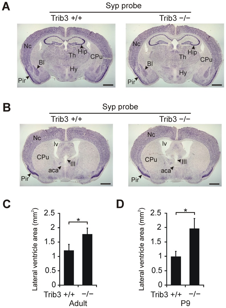
Figure 6. Gross brain morphology of Trib3 knockout (Trib3 −/−) and corresponding wild type (Trib3 +/+) mice.
(A and B) Representative adult mouse brain coronal sections are shown hybridized with a digoxigenin-labeled RNA probe complementary to mRNA encoding the synaptic vesicle protein Syp. (C) Size of lateral ventricles in adult Trib3 +/+ and Trib3 −/− mice (n = 7 per genotype). The area of the lateral ventricles was measured from coronal sections at the level depicted in panel B. (D) Size of lateral ventricles at postnatal day 9 (P9) in Trib3 knockout mice and their wild type littermates (n = 5 per genotype). In C and D, the areas of the left and right lateral ventricle on the coronal section were summed for each mouse, and the mean ± SEM for each genotype is presented. Abbreviations: aca, anterior commissure, anterior part; Bl, basolateral amygdala; CPu, caudate–putamen; Hip, hippocampus; Hy, hypothalamus; III, third ventricle; lv, lateral ventricle; Nc, neocortex; Pir, piriform cortex; Th, thalamus. Scale bar 1 mm. *P<0.05 comparing genotypes.

